

Dermatology
Microscopes for the dermatologist
Skin structure is the basis of a dermatologist’s examination of a patient’s skin. One of the most important tools for examining the skin is an illuminated magnifying glass or hand microscope. Where further assessment is required, fine tissue skin samples and data from biopsies are analysed using immunohistology tools, among other methods. In the following section you can find out which microscopes are suitable for this purpose. Magnifications from approximately 2x to 10x are common.
A more detailed analysis would then need to be carried out with (semi) plan apochromatic objectives, which are considerably more technically complex and reveal more detail, at magnification levels between 40x and 63x. If, for example, fungi and fungal spores are to be detected in untreated specimens, the use of fluorescent dyes is also an alternative to bright field microscopy for detection purposes.
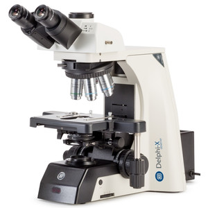
Euromex
Microscope DX.1153-PLPHi, phase, trino, infinity, 40x - 1000x
$ 8,100.00
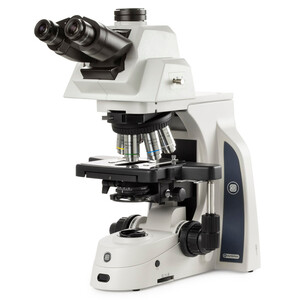
Euromex
Microscope Mikroskop DX.1158-APLi, trino, plan, apo, 40x-1000x, ergo head, AL, LED-3W
$ 11,800.00
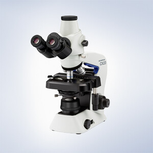
Evident Olympus
Microscope Olympus CX23 Photo, trino, plan, 40x,100x, 400x, LED
$ 2,080.00
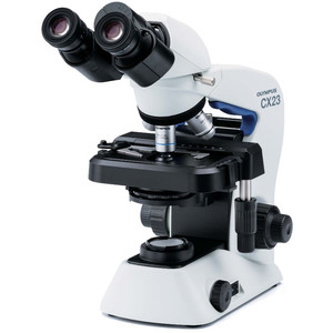
Evident Olympus
Microscope Olympus CX23 RFS1, bino, plan, achro, 40x,100x, 400x, 1000x, LED
$ 1,930.00

Evident Olympus
Microscope CX43 Standard, trino, infinity, LED, w.o. objectives!
$ 4,050.00
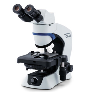
Evident Olympus
Microscope Olympus CX43 Ergo, bino, infinity, LED, w.o. objectives!
$ 5,300.00
