
Microscopes
Microscopy-Shop

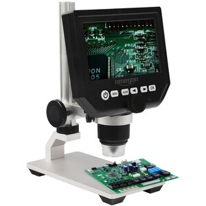
Micro-pro.com is online!
Visit our new online shop for microscopy and accessories! We offer a wide range of devices from all renowned manufacturers for various applications in science, industry, medicine, schools, education, and hobbies.
Concepts and features
Why are there such large price differences between different microscopes? A robust stand, high-quality illumination, colour-corrected optics - these are the things that matter.

A microscope contains several components that all deserve equal consideration.
- A robust, heavy stand that ensures a stable image and accurate focusing.
- High-quality illumination consisting of a light source, field diaphragm and condenser with aperture diaphragm.
- Colour-corrected optics, consisting of compatible eyepieces and lenses. In addition, importance should be attached to the microscope’s upgrade capabilities.
Why are there such large price differences between microscopes with the same magnification?
Magnification is no indication of quality. The difference comes from the stability of the mechanics, the high-quality optics with corrections for colour shift and edge sharpness. For example, up to 9 lenses may be required to correct edge sharpness. In addition, the light source is a quality criterion that ensures that the object is evenly illuminated. These three components must be perfectly matched to one another. Only in this way will the necessary contrast for a given magnification be achieved, which enables individual pixels to be distinguished.
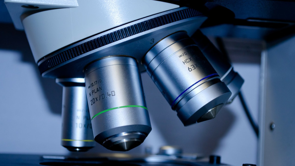
What does plan mean?
Plan means that the entire field of view is free of distortion, right up the edge. The curved lens makes the edge of the image distorted. This distortion is compensated for by the use of several counteracting lenses. Plan objectives play an important role especially in medical or routine applications.
For normal microscope use, chromatic correction is of greater importance. The objectives carry the designation achromatic, neofluar or apochromatic.

What does achromatic mean?
The refractive indices of different wavelengths (colours) are not identical. This leads to image defects, which are noticeable in the form of colour fringes around a specimen.
What is the difference between DIN and infinitely corrected optics?
In the case of DIN optics, the tube length is set at 160mm, the parfocal distance is 45mm and the objectives have uniform RMS threads. Theoretically, this standard means that optical microscope elements from different manufacturers can be combined. However, chromatic corrections are not homogeneous, which can lead to colour fringes around refractive objects.
With infinity-corrected optical systems, the effective tube length is extended by means of a parallel optical path inside the tube. Their main purpose is to allow the use of additional optical elements. An important example is the beam splitter, which is introduced into the optical path for photography. The light path is then focussed through a tube lens to produce an intermediate image. The focus lens is additionally used to correct chromatic aberration, resulting in a fully corrected intermediate image.
This is important for photography since chromatic correction is only carried out in the eyepiece of DIN optics, and a corrector lens is required in the case of trinocular microscopes. Another important field of application is fluorescence microscopy, where various filters are inserted into the tube. In addition to these benefits, the extended tube length minimises disruptive stray light. The disadvantage is that each manufacturer uses different components that are not interchangeable.
What is Köhler?

The Köhler illumination system consists of four components:
- a collector in the microscope’s foot
- a field diaphragm in the microscope’s foot
- condenser with
- aperture diaphragm
With Köhler illumination, these four elements are brought into focus in such a way that a parallel beam of light is spread evenly across the specimen.
Why do I need a field diaphragm?
The field diaphragm is used to limit the illuminated area to the field of view. This prevents disruptive stray light. It is also useful for centring the condenser.
Why do I need specimen protection?
The objective’s length increases with magnification. Especially with oil immersion objectives, the gap between the objective and the specimen is very small. As the objective is swung into place, the specimen can be damaged by the objective’s length. A spring mechanism in the objective tip prevents damage to the specimen.
What magnification do I need for my application?
10x - 40x
- minerals, stamps, electronics
- industrial applications
- insects, insect larvae
40x - 80x
- plant cells
40x– 100x
- larger single cells
100x– 200x
- fish parasites
- single cells (Vorticella)
400x
- fungi, pollen
- sperm
- blood
1000x
- bacteria
- chromosomes
Microscopes for various applications
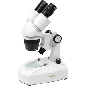
What's so special about stereo zoom microscopes?
Stereo microscopes tend to be overlooked when compared to biological microscopes, which sparkle with magnifications of up to 1000x.
Actually, they are the real magnification stars!
In contrast to other microscopes, stereo microscopes have two objective outputs and therefore guarantee stereo vision. Reflected and transmitted light ensure high contrast and depth of field, even with opaque specimens. Oblique illumination and shadowing enhance the 3D experience and make it easier to visualise the specimen. Laborious thin sections and preparations are not necessary. The large distance from the objective makes it easier to handle the specimen. A large field of view helps to keep a good overview.
Working with stereo zoom microscopes is particularly convenient: this is due to the usual variable magnification of between 7 and 45 times. Thanks to parfocal optics, only a small amount of focus adjustment is required when zooming. In premium devices, click-stops allow the measurement of specimen size. The appropriate exchangeable objective can double this magnification and thus 90-times magnification can be achieved, which already rules out free-hand manipulation of the specimen. However, a reduction of the magnification is also possible by means of an exchangeable objective, whereby the working distance increases.
Which stereo zoom microscopes are suitable for education, industry and research?
Firstly, we take a look at devices specifically designed for education and the classroom
Their features include:
- compact stand
- integrated reflected and transmitted light
- solid appearance
- microscopes for every budget
- Available for every requirement and all levels of education
They are particularly suitable for the identification of articulated animals and worms as well as their larvae. These can be observed live or prepared for anatomical examination. Stereo zoom microscopes are also indispensable for the study of botany, in particular comparative morphology. Geologists-to-be use stereo zoom microscopes to compare fossils and minerals. There are an infinite number of possible applications. That's why there is also a large range to choose from.
In order to provide an overview, we have created a summary table under the heading education.
Devices that are specially designed for industry include the following features
Key features:
- heavy, often with overhanging stand and variable height adjustment for the examination of large specimens
- ESD-safe materials for SMD
- large working distance to enable the manipulation of objects under the microscope
- often only with incident light or without a light source, specific illumination of opaque specimens with a ring or cold light source
- rugged construction, for daily use
Goldsmith or dental laboratory detailed work has long been unthinkable without a stereo microscope. But there are also countless applications for stereo zoom microscopes in modern manufacturing technology. Ever smaller and finer components require ever better image resolutions. This is especially true in the semiconductor and printed circuit board industries. Quality control and the documentation of component parts and their manufacturing processes are becoming increasingly important. Good optical devices with image data processing are essential for this. And can anyone imagine today’s forensic examination without a microscope and stereo microscope? In television crime series close to 100% of crimes are solved. How would this be possible without a stereo zoom microscope!
What are in-vitro diagnostics?
In-vitro diagnostic devices are devices and materials that comply with the German Medical Devices Act (MPG).
Extract from the Medical Devices Act (MPG) paragraph §3 from the German Federal Ministry of Justice and Consumer Protection.
Paragraph §3 Definition of terms (MPG)
In-vitro diagnostic device is a medical product which is used as a reagent, a reagent product, calibration material, control material, kit, instrument, apparatus, device or system, individually or combined, in accordance with the manufacturer’s defined purpose, is intended for in-vitro testing of samples obtained from the human body, including blood and tissue samples, and is intended solely or principally to provide information
- about physiological or pathological conditions or
- about congenital abnormalities or
- for testing the harmlessness to or tolerance by a potential recipient or
- for monitoring therapeutic measures.
Paragraph §4a quality assurance in medical laboratories
(1) Anyone carrying out medical tests in a laboratory setting shall establish a quality assurance system in accordance with the generally recognised medical science and technology standards in order to ensure the required quality, safety and performance in the use of in-vitro diagnostic devices and to ensure the reliability of the results thereby obtained. A proper quality assurance for medical laboratories is assumed if parts A and B1 of the guidelines of the Federal Medical Association for quality assurance in medical laboratory examinations of 23 November 2007 (German Medical Journal 105, pp. 341 to 355) are observed.
Paragraph § 31 medical product consultants
(1) Anyone who professionally informs or instructs other professionals on the proper handling of medical products (medical product consultants) may only carry out this activity if he has the necessary expertise and experience of the respective product and, if necessary, expertise in the instruction in the handling of the respective medical product. This also applies to information given by telephone.
(2) Has the expertise, has successfully completed a qualification in a scientific, medical or technical profession and has been trained on the respective medical devices.
Recommended microscopes