

Olympus CKX53
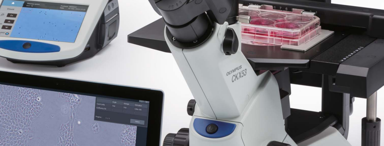
Compact, Ergonomic Inverted Microscope for Cell Culture
With improved image quality and ergonomics, the Olympus CKX53 offers reliable performance and an efficient, convenient workflow for the widest range of cell culture tasks; including live-cell observations, sample preparation, imaging, and fluorescence microscopy.
Evident Olympus
Inverted microscope Olympus CKX53 IPC/IVC V1, PH, trino, infinity, achro, 10x, 20x, 40x, LED
$ 8,500.00
Evident Olympus
CKX53 trinocular microscope, 100X, 200X, 400X, IPC / IVC x/y stage
$ 10,600.00
Evident Olympus
Inverted microscope Olympus CKX53 Hellfeld V2, trino, infinity, plan, achro, 2x, 4x, 10x, LED
$ 7,600.00

Evident Olympus
Inverted microscope Olympus CKX53 Hellfeld V1, trino, 40x, 100x,
$ 5,900.00
CKX53
Live cell microscopy
- Fast and efficient cell examination with integrated phase contrast (iPC) system
- Clear and wide visual field with 2X objective and FN 22
- Experience 3D vision with inversion contrast (IVC) technology
User-oriented design for efficient cell sampling and easy handling
- Efficient cell examination under sterile conditions
- Simple cell sampling in sterile workbench conditions
- Ergonomic, user-friendly design
- Suitable for a wide range of cell culture containers
- Better observation of multi-layer tissue culture flasks
- Flexible container selection
Objective
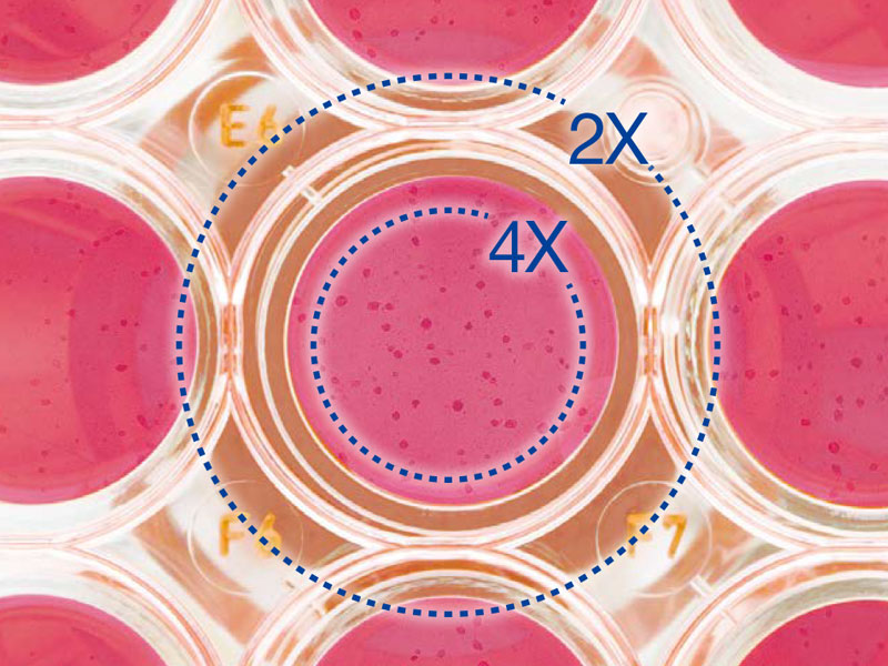
Efficient cell screening with a clear, wide field of view
- The PLN2X objective aperture diaphragm slider has a 22mm field of view and a diameter of 11 mm
- 2X objective provides noticeably higher contrast than other objectives. Even transparent objects in the sample can be clearly identified
- The wide field of view allows the detailed observation of individual cells in a 96-well plate without needing to move the stage
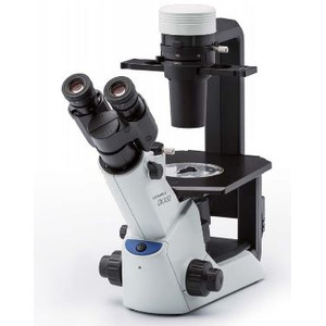
Ergonomics
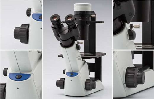
Ergonomic microscope design
Comfortable viewing position
The 45-degree angle of the eyepieces and the positioning of the butterfly-shaped observation tube against the stage enables ergonomic cell observation, whether standing or seated.
Convenient hand position close to the controls
All the controls, including the power switch, coarse and fine focus, and the knob for switching the light path, are ergonomically arranged for ease of operation and reduced user fatigue.
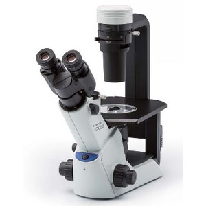
Comfort
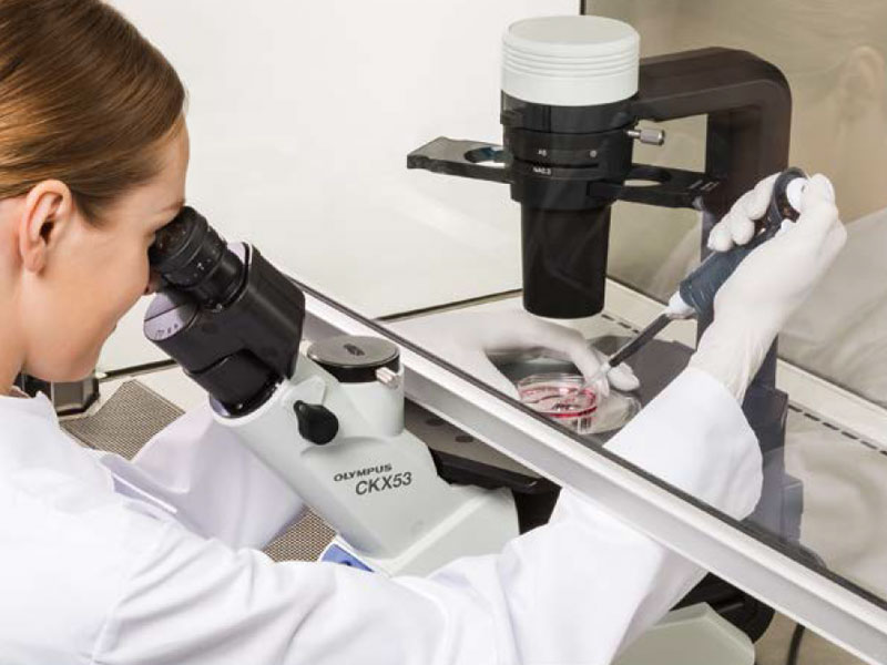
Observe live cells under sterile conditions
Small footprint enables sterilisation in the clean bench
The CKX53 microscope fits on a clean bench working surface and does not need to be removed during the UV sterilisation process thanks to its UV-resistant coating. The system is lighter than its predecessor, weighing around 7kg, and has a smaller footprint so it takes up less space in the laboratory. The microscope can be moved with one hand, and it’s easy to carry by the neck of the observation tube. The sliding pad in the base makes it easy to position.
Easy cell sampling in a sterile clean bench
The short distance between the viewing position and the optical axis and focus knob allows a natural hand position and eases focusing and cell sampling.
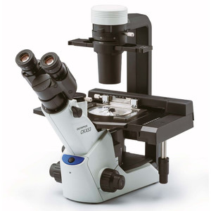
Optional specimen holder
Efficient cell observation and handling
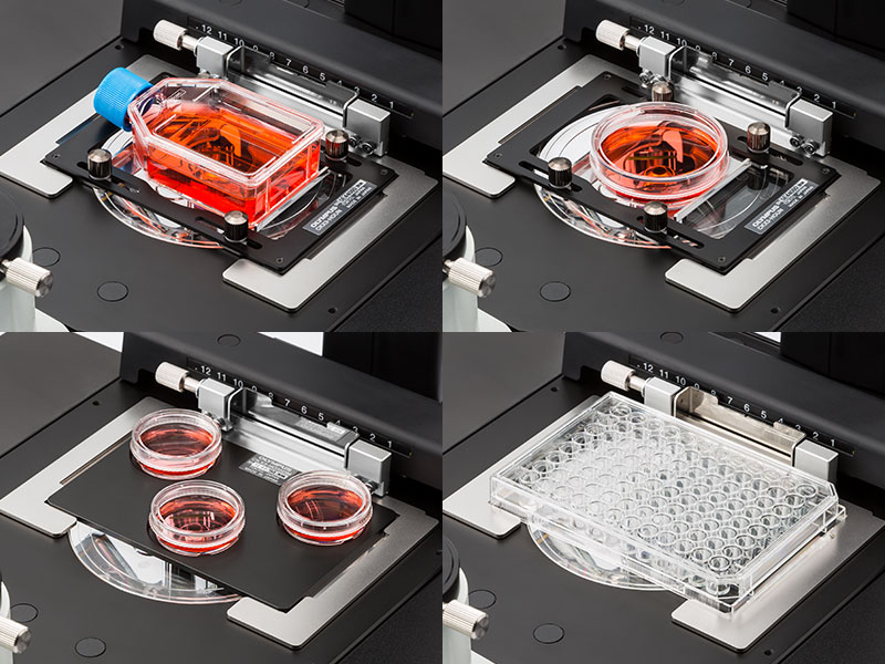
Suitable for a variety of cell culture containers
- The universal holder makes it easy to observe cells that were cultured in a variety of containers, including petri dishes, microplates, and flasks
- Three 35mm dishes fit on the stage with the optional holder, and various types of microplates can be used without the holder
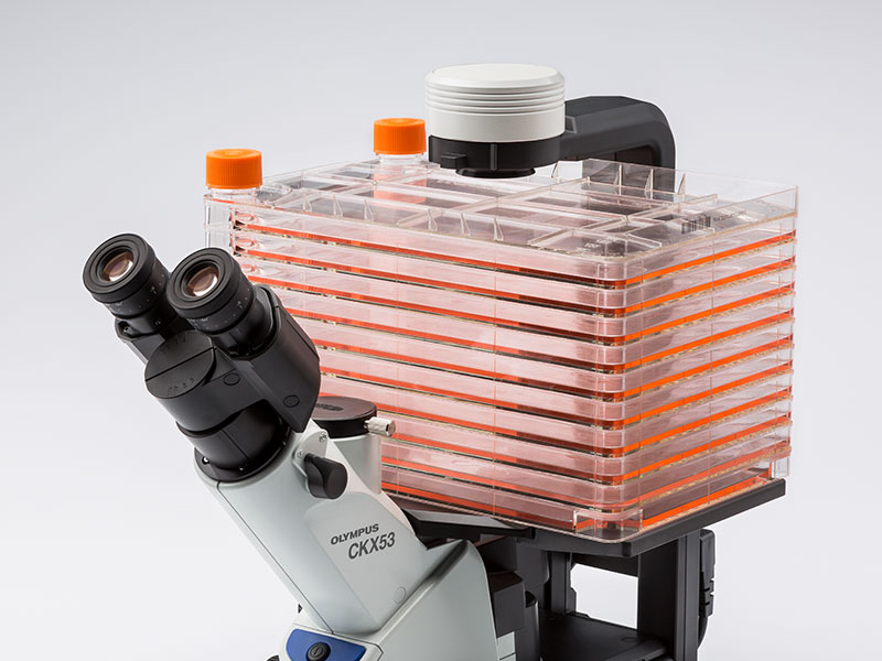
Comprehensive observation for a multilayer tissue flask
- The microscope's width and the easily detachable condenser enable the observation of containers, such as multilayer tissue flasks, up to 190 mm in height
- The PLCN4X objective’s depth of field allows cell observation of the two bottom layers of a multilayer flask
Also flexible enough for larger cell culture containers
- Movable holder for manually positioning culture containers
- Stage can be extended by up to 70mm to the left and right for greater flexibility
Integrated phase contrast system (iPC)
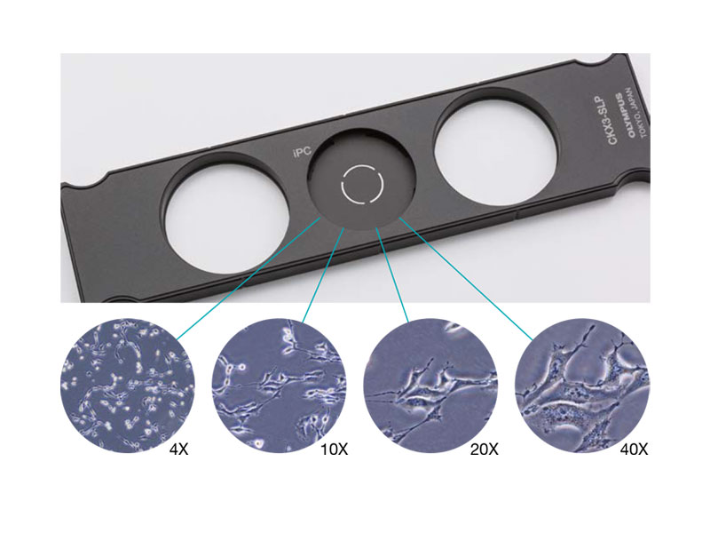
Simple observation of live cells
Fast observation of cells with the integrated phase contrast (iPC) system
- High contrast provides a clear view of cells at 4x, 10x, 20x, and 40x without needing to change or re-centre the ring slit
- The phase contrast system aids simple and efficient cell observation for a faster, more comfortable workflow
Inversion contrast (IVC)
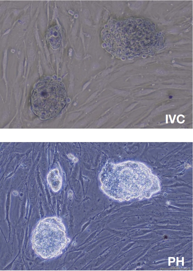
3D views of cells with the inversion contrast (IVC) technique
The IVC technique offers a reduced depth of field compared to phase contrast. This makes clear three-dimensional images for objects of any shape or transparency possible. IVC microscopy also provides clear views without halos or directional shadows, retaining the integrity of object details during observation.
10X objectives (PLCN10X, CACHN10XIPC) are used for this new IVC technique.
Image source: Y. Suzuki et al., Method for observing phase objects without halos or directional shadows. Opt Lett. 2015; 40(5): 812-5
Fluorescence
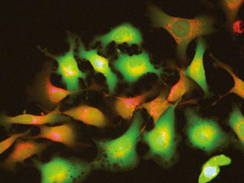
Easy fluorescence imaging
Clear images with a broad selection of fluorescent dyes
With the CKX53 standard fluorescence set, even weak fluorescence signals can be viewed clearly thanks to a range of integrable light sources, such as a 100W mercury lamp (U-LH100HG), a 130W high-pressure mercury lamp (U-HGLGPS), as well as 3rd party LEDs (not available in all locations). The filter holder's three slots accommodate the same type of filter module as used with the IX3 and BX3 microscopes.
High contrast under bright conditions
The umbra shield blocks ambient light effectively, enhances fluorescence contrast, and enables clear fluorescence observation under bright laboratory conditions. When using phase contrast, the umbra shield can be raised to allow light to pass through to the sample.
Further specifications
Dimensions (W × D × H): 200mm × 498mm × 454mm (phase contrast starting configuration)
Weight: ca. 6.9kg
Objectives
Efficient Cell Screening with a Clear, Wide View
- Efficiently screen for desired cells thanks to the ring slit for the PLN2X objective, which offers a 22 mm field of view and an 11 mm diameter
- 2X objective provides noticeably higher contrast than other objectives, so even transparent objects in the sample can be clearly identified
- Wide visual field makes it easy to observe the cells in individual wells of a 96-well plate without moving the stage
Ergonomy
Ergonomic Microscope Design
Comfortable Viewing Position
The eyepieces’ 45-degree angle and the placement of the butterfly-shaped observation tube against the stage facilitates ergonomic cell observation, whether standing or seated.
Rest Your Hand Near the Controls
All the controls, including the power switch, coarse and fine focus, and the knob for switching the light path, are ergonomically located for easy access and reduced user fatigue.
Comfort
Live Cell Observation Under Sterile Conditions
Small Footprint Enables In-Hood Sterilization
The CKX53 microscope fits on a clean lab bench and can be remain there during the UV sterilization process thanks to its UV-resistant coating. The system is about 7 kg (15.4 lb), lighter than the previous model and has a smaller footprint, so it takes up less space in your lab. You can move the microscope with one hand, and it’s easy to carry it by the neck of the observation tube. The base’s sliding pad makes it easy to position.
Easy Cell Sampling in a Sterile Bench Environment
The short distance between the view point and the optical axis/focus knob facilitates natural hand positioning and makes focusing and cell sampling easier.
optional x/y-stage
Efficient Cell Observation and Handling
Accommodates a Variety of Cell Culture Containers
- The universal holder makes it easy to view cells that were cultured in a variety of containers, including dishes, microplates, and flasks
- Three 35 mm dishes can fit on the stage with the optional holder attachment
- Different types of microplates can be accommodated without the holder
Comprehensive Observation for a Multilayer Tissue Flask
- The microscope’s width and easily detachable condenser enable you to view containers, such as multilayer tissue flasks, up to 190 mm (7.5 in.) in height
- The PLCN4X objective’s depth of focus makes it easy to observe cells in the two bottom layers of a multilayer flask
Flexibility to Use Larger Cell Culture Containers
- Lift the holder’s arm to manually position culture containers
- Expand the stage up to 70 mm (2.8 in.) to the left and right for greater handling flexibility
Integrated Phase Contrast (iPC)
Easy Live Cell Observations Fast
Cell Observation with the Integrated Phase Contrast (iPC) System
- High contrast provides a clear view of cells at 4x, 10x, 20x, and 40x without the need to exchange or re-center the ring slit
- Phase contrast system facilitates simple and efficient cell observation for a faster, more comfortable workflow
Inversion Contrast (IVC)
3D Cell Views with the Inversion Contrast (IVC) Technique
The IVC technique offers a narrower depth of field than phase contrast, rendering clear three-dimensional images for objects of any shape or transparency. IVC observation also provides clear views without halos or directional shadows, preserving the integrity of object details during observation.
10X objectives (PLCN10X, CACHN10XIPC) are used for this new IVC observation.
Reference: Y. Suzuki et al., Method for observing phase objects without halos or directional shadows. Opt Lett. 2015; 40(5): 812-5Fluorescence
Easy Fluorescence Imaging
Clear Views with a Wide Range of Fluorescent Dyes
With the CKX53 standard fluorescence set, even weak fluorescence signals can be viewed clearly with the aid of different integrated light sources, such as a 100 W mercury lamp (U-LH100HG), a LED and LDP light source (U-LGPS), and 3rd party LEDs (Not available in some areas). The same type of mirror unit as those offered with our IX3 and BX3 microscopes can be set in the mirror unit slider’s three slots.
High Contrast Under Bright Conditions
The Umbra Shield efficiently blocks out room light, enhances the contrast of fluorescence, and enables clear fluorescence observation under bright lab conditions. When using phase contrast, you can lift the shield to pass light through to the sample.
Further Specifications
Dimensions (W × D × H): 200 mm × 498 mm × 454 mm (phasecontrast standard configuration)
Weight: Approx. 6.9 kg